aEEG Background Patterns
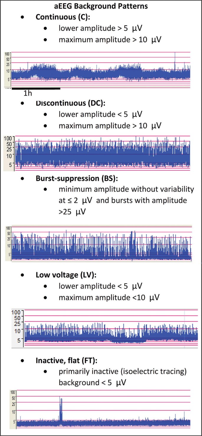
Fig. 16.2 — Common classification used to describe aEEG tracing, especially in term infants: continuous (C) with SWC, discontinuous (DC), burst suppression (BS), low voltage (LV), and inactive, flat trace (FT).
Cite this chapter
El-Dib, M., de Vries, L.S. (2024). Neurophysiological Monitoring. In: Meijler, G., Mohammad, K. (eds) Neonatal Brain Injury. Springer, Cham. https://doi.org/10.1007/978-3-031-55972-3_16
Open Access, licensed under CC BY 4.0. Figure reproduced with attribution; no changes made.
Open Access, licensed under CC BY 4.0. Figure reproduced with attribution; no changes made.